Agarose_Gel_Electrophoresis

Agarose Gel Electrophoresis
琼脂糖凝胶电泳
三、试剂与器材(Reagents and apparatus)
1. Agarose.
2. TBE [5 × stock solution (1 liter): 54 g Tris base, 27.5 g boric acid, 20ml 0.5 M EDTA, pH 8.0].实验室:TAE50×
3. 10 × loading buffer: 0.25% bromophenol blue, 40% sucrose in water.
实验室:碧云天 6×
4. Equipment: beaker, graduated cylinder, stir bar, microwave, Pan balance, comb, electrophoresis tank, and Electro-phoresis System , Ultraviolet transilluminator.
5. EB实验室:分装5mg/mL
四、实验步骤(Experimental Procedures)
Ⅰ. Preparation of the gel (凝胶的制备)
1.制备1%琼脂糖凝胶。称取0.5g琼脂糖,放人锥形瓶中,加入50mL的 1×TAE 缓冲液,放入微波炉加热至完全溶化,则为1%琼脂糖凝胶液。(由于蒸发作用,溶解前在容量瓶上作一个记号或称量质量,溶解后用三蒸水补足)
2.制胶器的安装
①取多功能制胶器,洗净,晾干;
②将多功能制胶器放置于一水平位置,选择12×6cm制胶架,然后选择1.5mm 18teeth的梳子(最大加样量25μl);
③加入电泳缓冲液(1×)至电泳槽中
3. 将熔化的琼脂糖凝胶液转入三角瓶中,然后加入EB 5μl。
4. 将冷到60℃左右的琼脂糖凝胶液,缓缓倒入所选择的制胶槽内,直至有机玻璃板上形成一层均匀的胶面(注意不要形成气泡)。
5. 将所选择规格的梳子插入制胶架的定位槽中。
6. 待胶凝固后(30-60min),轻轻拔掉梳子,将凝胶盘从制胶槽中取出,放入电泳槽内。
Ⅱ. Loading DNA samples (加样)
用移液枪缓慢将DNA样品垂直加入加样孔直至开口下方。
1.DNA samples :4-5μl
2. Loading buffer
(6X):1μl(用移液枪滴于EP手套上,与DNA samples来回吹打混匀)
3. DNA markers :6μl
Ⅲ.Gel(电泳)
1. 接通电泳槽与电泳仪的电源(注意正负极,DNA片段从负极向正极移动)。保持电压 60-80 V。实验室:120V左右
2. 当溴酚蓝染料移动到距凝胶前沿1-2cm处,停止电泳。(看情况而定)
Ⅳ. Gel Interpretation (凝胶图像解释)
The uncut DNA lane may have several bands in it. This occurs because the mobility of plasmid DNA in an agarose gel depends on its molecular conformation as well as its size in base pairs. Plasmid DNA can exist in any one of three major conformations:
未切割的质粒DNA在其泳道上也许会出现几个条带,之所以这样是由于质粒DNA在琼脂糖凝胶中的迁移距离是由其分子构象及其碱基对大小所决定的。质粒DNA以下列三种主要构象中的任何一种形式存在:
Supercoiled -- plasmid is usually seen as a supercoil in a bacterial cell. This form of the plasmid will move the fastest through the gel due to its compact structure. 超螺旋——质粒在细菌细胞以超螺旋结构存在,由于其结构致密,它在凝胶中的泳动速度最快。 Nicked--During plasmid DNA replication, topoisomer-ase I introduces a nick into one
strand of the DNA helix and uncoils the plasmid. Physical shearing and enzymatic cleavage during plasmid isolation may also introduce nicks into the supercoiled plasmid to produce a relaxed open circular structure.
切口——在质粒DNA 复制过程中,拓扑异构酶I 会在DNA 双螺旋中的一条链中引入一个切口,解开质粒的超螺旋。在质粒分离过程中由于物理剪切和酶的切割作用同样也会在超螺旋质粒中引入切口从而产生松散的开环结构。这种形式的质粒迁移速率介于超螺旋和线性DNA 之间。 Linear ——Linear plasmid DNA occurs when damage results in strand nicks directly opposite each other on the DNA helix. This form is the slowest migrating form of plasmid. The presence of linear DNA in a plasmid preparation is a sign of either nuclease contamination or sloppy lab procedure.
线性——当DNA 损伤在DNA 双链相对应的两条链上同时产生切口时,就会出现线性质粒DNA ,这种DNA 的泳动速率最慢,质粒制备过程个出现线性DNA 说明存在核酸酶污染或实验操作有问题。
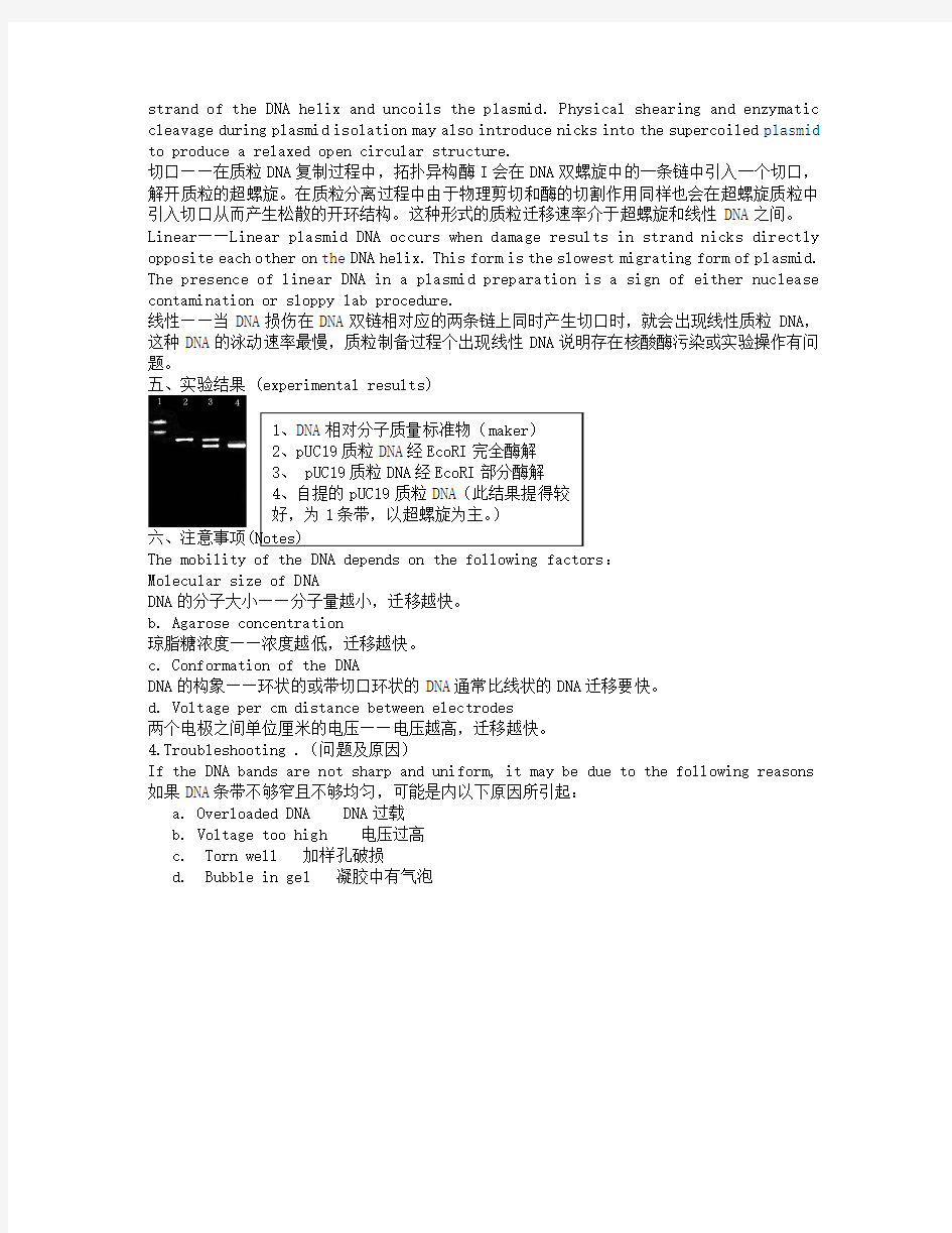
五、实验结果 (experimental results)
六、注意事项
The mobility of the DNA depends on the following factors :
Molecular size of DNA
DNA 的分子大小——分子量越小,迁移越快。
b. Agarose concentration
琼脂糖浓度——浓度越低,迁移越快。
c. Conformation of the DNA
DNA 的构象——环状的或带切口环状的DNA 通常比线状的DNA 迁移要快。
d. Voltage per cm distance between electrodes
两个电极之间单位厘米的电压——电压越高,迁移越快。
4.Troubleshooting .(问题及原因)
If the DNA bands are not sharp and uniform, it may be due to the following reasons 如果DNA 条带不够窄且不够均匀,可能是内以下原因所引起:
a. Overloaded DNA DNA 过载
b. Voltage too high 电压过高
c. Torn well 加样孔破损
d. Bubble in gel 凝胶中有气泡