PDF (212.8 KB)
Introduction
Glaucoma is a chronic neurodegenerative disease in which apoptosis of retinal ganglion cells and progressive loss of optic nerve axons eventually leads to blindness.1,2 Elevation of intraocular pressure (IOP), ocular hypertension, is a major risk factor in the most prevalent type of glaucoma, primary open-angle glaucoma (POAG). However, the mechanism of neural damage by ocular hypertension is not completely understood. Ocular hypertension is induced by glucocorticoids, such as dexamethasone (DEX), in experi-mental animal models and a subset of patients treated sys-temically or topically (steroid-induced glaucoma).3,4 Ocular hypertension in both POAG and steroid-induced glaucoma results from an increased aqueous out? ow resistance across the trabecular meshwork (TM), and histopathological ? nd-ings of the TM in POAG and steroid-induced glaucoma share some similarities,5–7 although the mechanism for this increased resistance is also poorly understood.
Proteomic approaches play important roles in dissecting unknown molecular mechanisms of complex physiological/
Jpn J Ophthalmol 2008;52:84–90 DOI 10.1007/s10384-007-0507-5? Japanese Ophthalmological Society 2008
LABORATORY INVESTIGATION
Proteomic Analysis of Rat Retina in a Steroid-
Induced Ocular Hypertension Model: Potential
Vulnerability to Oxidative Stress
Nariko Miyara1,2, Manabu Shinzato1,2, Yoshito Yamashiro2, Akihiro Iwamatsu3,
Ken-ichi Kariya2, and Shoichi Sawaguchi1
1Department of Ophthalmology and Visual Sciences, School of Medicine, University of the
Ryukyus, Okinawa, Japan; 2Division of Cell Biology, Graduate School of Medicine, University
of the Ryukyus, Okinawa, Japan; 3Protein Research Network, Yokohama, Japan
Abstract
Purpose: To investigate global protein expression pro? les in the retinas of normal and glucocorticoid-
induced ocular hypertensive rats by proteomic analysis.
Methods: Ocular hypertension was induced by topical application of dexamethasone (DEX) for 4 weeks.
Age-matched untreated rats served as controls. Intraocular pressure (IOP) was monitored by an elec-
tronic tonometer. Retinal protein expression pro? ling was carried out by two-dimensional ? uorescence
difference gel electrophoresis (2-D DIGE). Proteins were identi? ed by matrix-assisted laser desorption
ionization time-of-? ight (MALDI-TOF) mass spectrometry.
Results: In DEX-treated rats, average IOP was elevated signi? cantly compared with controls. With DEX
treatment, levels of four proteins were altered, as revealed by 2-D DIGE and MALDI-TOF mass spec-
trometry: apolipoprotein A1 (apoA1), a lipid-binding protein, upregulated 1.9-fold, P< 0.05; alpha A
crystallin (CRYAA), a molecular chaperone, downregulated 2.7-fold, P< 0.01; superoxide dismutase 1
(SOD1), an antioxidant enzyme, downregulated 2.3-fold, P< 0.05; and triosephosphate isomerase 1
(TPI1), a glycolytic enzyme, downregulated 2.3-fold, P< 0.01.
Conclusions: Downregulation of CRYAA, SOD1, and TPI1, observed here after a short period of DEX-
induced ocular hypertension, may be involved in the onset of neural damage in steroid-induced
glaucoma. Jpn J Ophthalmol 2008;52:84–90 ? Japanese Ophthalmological Society 2008
Key Words:glucocorticoid, ocular hypertension, proteomics, retina
Received: July 9, 2007 / Accepted: September 17, 2007
Correspondence and reprint requests to: Ken-ichi Kariya, Division
of Cell Biology, Graduate School of Medicine, University of the
Ryukyus, 207 Uehara, Nishihara-cho, Okinawa 903-0215, Japan
e-mail: kariya@med.u-ryukyu.ac.jp
N. MIYARA ET AL. 85 RETINA PROTEOMICS OF A STEROID-INDUCED GLAUCOMA MODEL
pathological processes.8 For example, the strategy of cou-pling Ettan two-dimensional ?uorescence difference gel electrophoresis (2-D DIGE, GE Healthcare, Little Chalf-ont, Buckinghamshire, UK) with mass spectrometry has proven to be a powerful tool for proteomic analysis.9 The 2-D DIGE system is free from the limitations of traditional stain-based systems in two aspects: ?rst, proteins in the control and test samples are labeled with different ? uores-cent dyes and subjected to two-dimensional separation on the same gel, enabling perfect protein spot matching, and second, linear dynamic ranges for ? uorescent dyes have ? ve orders of magnitude, allowing detection of previously unde-tectable spots and very accurate quantitative comparisons. Protein spots exhibiting quantitative differences are subse-quently examined by mass spectrometry, and proteins con-tained in these spots can be identi? ed.
In a preceding study, we applied the above strategy to gain insight into the mechanism of glucocorticoid-induced ocular hypertension.10 We used a rat ocular hypertension model generated by relatively short-term topical DEX application,11 and compared global protein expression pro-? les in the TM of control and DEX-treated rat eyes.10 Type I collagen C propeptides, which are processing by-products of ? brillar collagen, were downregulated. The data strongly suggested that collagen turnover, a cycle of homeostatic degradation and repair by newly formed ? brils, is slowed in DEX-treated eyes. Consistent with this, there exists a mouse genetic model in which impaired turnover of TM type I collagen is associated with ocular hypertension.12 In the present study, we used the same strategy to examine retinas of DEX-induced ocular hypertensive rats. Apolipoprotein A1 (apoA1) was upregulated, and alpha A crystalline (CRYAA), superoxide dismutase 1 (SOD1, Cu/Zn SOD), and triosephosphate isomerase 1 (TPI1) were downregu-lated. As downregulation of optic nerve SOD1 and TPI1 has also been noted in a human proteomic study of POAG patients,13 the present results could be relevant to under-standing the mechanism of neural damage in ocular hyper-tensive eyes.
Materials and Methods
Rat Ocular Hypertension Model
Six male Wistar rats (weighing approximately 250 g) were purchased and divided into three pairs (rat 1 versus 4; rat 2 versus 5; rat 3 versus 6). Three rats (rats 4, 5, and 6) were treated with topical application of DEX on both eyes for 4 weeks to induce ocular hypertension as described previ-ously.10,11 The other rats (rats 1, 2, and 3) were treated with vehicle only and served as controls. At the end of treatment, they were used for experiments after IOP measurement conducted with an electronic tonometer as described.10,11 All experiments were conducted in compliance with the University of the Ryukyus Animal Care and Use Commit-tee Guidelines and the ARVO Statement for the Use of Animals in Vision Research.Protein Extraction and Fluorescent Dye Labeling
Both eyes of each rat were gently enucleated. The neuro-sensory retina was separated from the underlying retinal pigment epithelium layer, washed in ice-cold phosphate-buffered saline, and collected in 150 μl of homogenization buffer [2 M thiourea, 7 M urea, 4% w/v 3-[(3-cholamido-propyl) dimethylammonio]-1- propanesulfonate (CHAPS), 30 mM Tris-HCl, pH 8.5], supplemented with Complete EDTA-free Protease Inhibitors (Roche Applied Science, Penzberg, Germany). After homogenization with Pellet Mixer (TreffLab, Degersheim, Switzerland) and a probe sonicator, the crude homogenate was centrifuged at 16 000 g for 30 min at 4°C. Proteins in the supernatant were puri? ed with the 2-D Clean Up kit (GE Healthcare) and redissolved in homogenization buffer. Protein concentration was deter-mined using the 2-D Quant kit (GE Healthcare) and adjusted to 5 μg/μl, before the proteins were stored at ?80°C. After thawing on ice and centrifugation at 16 000 g for 10 min at 4°C, proteins were labeled with CyDye DIGE ?uorescent dyes according to the manufacturer’s instruc-tions (minimal labeling protocol, GE Healthcare). Control and DEX-treated samples were differentially labeled with Cy3 and Cy5, respectively, or vice versa.
Two-Dimensional Separation of Labeled Proteins and Image Analysis
For the ?rst dimension of isoelectric focusing (IEF), an immobilized pH gradient (IPG) dry strip was used (pH 4–7, 18-cm linear, Immobiline DryStrips, GE Healthcare). A pair of labeled control and DEX-treated samples (50 μg total proteins each) were mixed (total 24 μl), combined with 320 μl of rehydration buffer [8 M urea, 2% CHAPS, 0.002% w/v bromophenol blue (BPB), 0.5% v/v IPG buffer (GE Healthcare) and 1.2% v/v Destreak Reagent (GE Health-care)] and applied to the dry strip. After overnight rehydra-tion loading (in gel rehydration) of sample proteins, the strip was subjected to IEF with the LKB2117 Multiphor II (GE Healthcare) and a programmed voltage gradient (0 to 500 V ramp for 1 min, 500 to 3500 V ramp for 1.5 h, and 3500 V for 6 h).
For the second dimension of sodium dodecyl sulfate-polyacrylamide gel electrophoresis (SDS-PAGE), the strip after IEF was equilibrated for 15 min in equilibration buffer (50 mM Tris-HCl, pH 8.8, 6 M urea, 30% v/v glycerol, 2% w/v SDS, 0.002% BPB and 1% w/v dithiothreitol). The strip was then mounted on top of a 12.5% gel (1 mm thick, 24 cm × 24 cm) that was cast using low-? uorescence glass plates (Nihon EIDO, Tokyo, Japan) and ?xed in place with a shark-tooth comb. SDS-PAGE was carried out overnight at a constant temperature (20°C) and constant power (60 mA) until the BPB dye front was approximately 1 mm from the bottom of the gel. The gel was scanned by using a Typhoon Variable Mode Imager (GE Healthcare) to acquire Cy3 and Cy5 images. Protein spots from each image were outlined and quanti? ed with Image Quant Tool and Image Master
86 Jpn J Ophthalmol
Vol 52: 84–90, 2008 Platinum software programs (GE Healthcare). The quan-
tity of the individual spot in each image was expressed as
percent volume (%V), relative to the sum of all detected
spots from each image.14
Screening and Identi? cation of Differentially
Expressed Proteins
For screening of protein spots altered by DEX treatment,
three pairs of control and DEX-treated rat samples (rat 1
versus 4; rat 2 versus 5; rat 3 versus 6) were analyzed as
described above. In two analyses (rat 1 versus 4; rat 3 versus
6), control and DEX-treated samples were labeled with Cy3
and Cy5, respectively. In one analysis (rat 2 versus 5), the
labels were reversed, as we have found a few proteins where
Cy3 labeling proved more ef? cient than Cy5, as well as the
opposite (data not shown). Only protein spots that ex-
hibited similar DEX-induced changes in all three analyses
were selected. Using the %V value, a ratio of DEX-
induced change in each selected spot (%V in DEX-treated
sample/%V in control sample) was then calculated for each
of the three analyses. Finally, the average ratio of all three
analyses was calculated. Only protein spots that exhibited
statistically signi? cant changes in the average ratio (P< 0.05
or P< 0.01 by paired t test for three pairs) were de? ned as altered by DEX treatment and subjected to protein identi-?cation. Most protein spots were unaltered: correlation coef? cients of spot intensities (%V values) in all three pairs of control and DEX-treated rat samples were >0.9.
For identi?cation of proteins, which requires a large amount of proteins, 25 μg of a Cy3-labeled sample from rat 3 was combined with Cy5-labeled samples from all other rats (total protein, 500 μg). After two-dimensional separa-tion, proteins in the gels were transferred to polyvinylidene di?uoride (PVDF) membrane (ProBlott PVDF, Applied Biosystems, Foster City, CA, USA) followed by Colloidal Gold Total Protein staining (BioRad Laboratories, Hercu-les, CA, USA). Protein spots altered by DEX treatment were excised (1 mm × 1 mm square) from the membrane. Immobilized proteins were reduced, S-carboxymethylated, and digested in situ with Achromobacter protease I (a Lys-C), which cleaves after Lys residues.15 Molecular mass anal-yses of Lys-C fragments were performed by matrix-assisted laser desorption/ionization time-of-? ight (MALDI-TOF) mass spectrometry, using the Applied Biosystems Voyager-DE/STR mass spectrometer.16 Identi? cation of proteins was carried out by peptide mass ?ngerprinting and screening using the National Center for Biotechnology Information non-redundant protein database (http://www.ncbi.nlm.nih. gov/). A mass tolerance of ±0.03 Da was used for the peptide mass ? ngerprinting.
Results
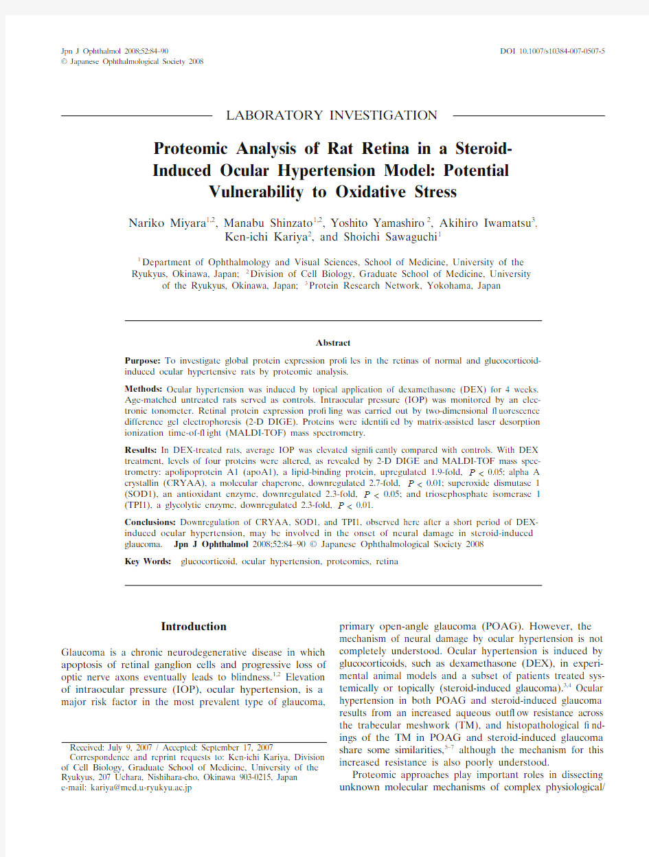
Glucocorticoid-induced ocular hypertensive rats were gen-erated according to the protocol of our previous studies.10,11IOPs of rats treated for 4 weeks with topical DEX were signi? cantly elevated compared with those of age-matched, vehicle-treated, control rats (P< 0.05; Fig. 1). IOPs of control and DEX-treated rats were also comparable to those in our previous studies.10,11
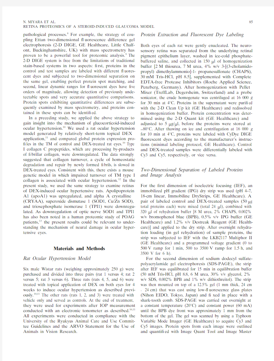
To understand the mechanism of retinal damage in DEX-induced ocular hypertension, we sought to identify by using a proteomics approach retinal proteins whose expres-sion levels were altered by DEX treatment. To this end, we extracted retinal proteins from control and DEX-treated rat eyes and examined global protein expression pro? les using 2-D DIGE, a powerful two-dimensional differential gel electrophoresis system (Fig. 2). In this system, different protein samples are labeled with different ? uorescent dyes and mixed. Mixed labeled proteins are separated in the same gel, according to their isoelectric point (pI) in the ? rst dimension and to their size in the second dimension. Sepa-rated proteins are represented by ? uorescent spots on the gel, which are visualized by capturing ? uorescence images of the gel for each dye. Using this system, we analyzed protein expression pro?les in three pairs of control and DEX-treated rat retina samples. We found four protein spots (out of 900–1000 spots) with intensities consistently altered by DEX treatment in all three analyses (designated spots 1 to 4).
These protein spots exhibited various degrees of DEX-induced alterations as revealed by pseudocolor representa-tion (Fig. 2) and quantitative calculations (Table 1). First, the intensity of each spot in each analysis was expressed as %V, where volume (integrated area and ? uorescence strength) of each spot was normalized to the sum of volume of all the spots in the gel. Second, %V of each spot in DEX-treated samples was normalized to that of control samples 16
18
20
22
24
26
28
30
32
34
P
o
s
t
-
t
r
e
a
t
m
e
n
t
I
O
P
(
m
m
H
g
)
Control
(n=3)
DEX
(n=3)
Figure1.Intraocular pressure (IOP) (circles) in rats after 4 weeks of treatment (topical administration, four times daily) with vehicle only (control) or dexamethasone (DEX). n, number of rats. *P< 0.05.
N. MIYARA ET AL.
87
RETINA PROTEOMICS OF A STEROID-INDUCED GLAUCOMA MODEL
to obtain the ratio of DEX-induced change. Finally, the ratios in three analyses were averaged to give the average ratio. Among the four protein spots, the average ratio varied from 0.430 to 1.920, and all changes were statistically signi? cant (P < 0.05 for spots 1 and 3, P < 0.01 for spots 2 and 4 by paired t test; Table 1). Thus, these four spots were de? ned as having been altered by DEX treatment. Of these four spots, one was upregulated by DEX treatment (spot 1), while the others were downregulated (spots 2–4).We successfully identi? ed proteins contained in all of these spots. In 2-D DIGE, protein spots visualized with ? uorescence scanning do not always indicate the highest point of protein concentration on the gel.9 Most protein spots contain labeled as well as unlabeled protein: in our minimal labeling procedure only a small fraction of proteins were ? uorescence-labeled.9 Fluorescence-labeling causes retarded migration of proteins in the second dimension, an effect more marked for small proteins.9 In order to subject the area of highest protein concentration to the protein identi? cation procedures, we electroblotted protein spots to PVDF membranes, visualized them by colloidal gold total protein staining, and excised them. Transfer of protein spots from the gel to a membrane also allowed ef? cient reduction and S-alkylation of disul? de bonds and digestion with Lys-C protease (on-membrane digestion). We obtained peptide mass spectra by MALDI-TOF mass spectrometry, and identi? ed proteins using two complementary and con-? rmatory engines for bioinformatics database searching (Mascot Search and Profound Search, peptide mass ? ngerprinting).The identi? ed altered proteins included (protein name abbreviation, spot number, and DEX-induced change, in parentheses): apolipoprotein A1 (apoA1, spot 1, upregu-lated 1.9-fold); alpha A crystallin (CRYAA, spot 2, down-regulated 2.7-fold); superoxide dismutase 1 (SOD1, spot 3, downregulated 2.3-fold); triosephosphate isomerase 1 (TPI1, spot 4, downregulated 2.3-fold). Their database accession numbers, theoretical molecular weights and pI, total amino acid numbers, matched peptides (residue numbers), and percentage of coverage of protein sequence (% coverage, matched amino acid/total amino acid × 100) are shown in Table 2.
A
M o l e c u l a r m a s s (k D a )
116
66453121
14
B
4
1
Figure 2A, B. Two-dimensional ? uorescence difference gel electro-phoresis (2-D DIGE). Retinal protein samples from control and DEX-treated rat eyes (50 μg protein each) were labeled with Cy3 and Cy5 ? uorescent dyes, respectively, or vice versa. Samples were mixed and separated in the same gel on the basis of pI (x axis) and molecular mass (y axis). Three analyses using independent pairs of control and DEX-treated samples gave essentially the same results. A Overview of the control Cy3 ? uorescent image (grayscale image for Cy3 channel). B. Magni? ed image of the boxed region in A . Arrows with numbers indi-cate protein spots with intensities that were consistently altered by DEX treatment in all three analyses. Merged pseudocolor images are also presented: green for Cy3 and red for Cy5 channels. Protein spots upregulated or downregulated by DEX treatment appear red or green, respectively, while unaltered protein spots appear yellow.
Table 1. Spot intensities of control and DEX-treated samples
Spot no.a Control %V b
DEX %V b
Average ratio c
P value d 10.02150.0413 1.9200.03120.03090.01130.3670.00730.00490.00220.4400.0354
0.0121
0.0052
0.430
0.002
DEX, dexamethasone.a
Spot numbers correspond to those of the four protein spots in Fig. 2.b
Intensities of each spot in control and DEX-treated samples were expressed as percent volume (%V), and %V of three independent analyses were averaged.c
The ratio of spot intensity of the DEX-treated sample to that of the control sample (DEX-treated/control) was calculated in each of three independent analyses and averaged.d
Paired t test of three pairs.
88 Jpn J Ophthalmol
Vol 52: 84–90, 2008
Discussion
To our knowledge, this is the ?rst quantitative proteomic analysis of ocular hypertensive retina. The only precedent of proteomic analysis of ocular hypertensive retina is a study by Tezel et al.,17 who examined oxidative modi? ca-tion, rather than expression level, of retinal proteins. Nev-ertheless, their study is relevant to ours, as discussed later. In their study, IOP elevation of approximately 10 mmHg was attained in rats by injections of hypertonic saline into episcleral veins. Judging from the ?gures in their paper, they probably extracted retinal proteins after >7 weeks of ocular hypertension. Traditional two-dimensional electro-phoresis gave >400 spots. Of these >400 spots, 60 exhibited protein carbonyl immunoreactivity, which re? ects oxidative modi?cation, in ocular hypertensive retinas. Of these 60 spots, 20 were found modi? ed only in ocular hypertensive retinas, whereas the rest were also modi?ed in control retinas, albeit to a signi? cantly lesser extent. Three major modi?ed proteins speci?c to ocular hypertensive retina were identi?ed by mass spectrometry: glyceraldehyde-3-phosphate dehydrogenase (GAPDH), a glycolytic enzyme; heat shock protein 72 (HSP72, also known as HSP70), a molecular chaperone; and glutamine synthetase, an excito-toxicity-related protein.17 In the present study, 2-D DIGE showed >900 spots, but only four (apoA1, CRYAA, SOD1, and TPI1) were altered by ocular hypertension. Although ocular hypertension may affect mainly protein modi? ca-tions rather than protein expression levels, this result might also be attributed to a mild and short-term ocular hyperten-sion in our model. In our DEX-induced model, statistically signi?cant IOP elevation of approximately 5 mmHg was attained only after 2 weeks of treatment.11 Elevation of approximately 10 mmHg was attained only at the end of 4 weeks of treatment. Thus, the present results may re? ect etiologically important primary changes at very early stages of retinal damage, which in later stages are dif? cult to dis-tinguish from many other secondary changes. In this regard, it is interesting to note that SOD1 and TPI1 were among 32 downregulated proteins in a human proteomic study of POAG optic nerve13 and that the CRYAA gene was among
36 downregulated retinal genes in a microarray analysis of
a DBA/2J mouse glaucoma model.18
Downregulation of SOD1 might cause vulnerability to oxidative stress. Elevated oxidative stress has been reported repeatedly in ocular hypertensive retinas.19,20 In addition to
Table2. Protein identi? cation by mass spectrometry
Spot no.a
Protein name
(accession no.)b
Molecular
mass (Da)c pI d
Total amino
acid residues e
Matched peptides
(residue nos.)f
Percent
coverage g
1Apolipoprotein A-I
(NP_0.36870.1)30062 5.73 25936–46
47–63
101–111
120–130
131–141
142–151
155–161
182–201
202–212
224–243
246–254
246–255
55
2Alpha A crystallin
(NP_036666.2)19792 6.2017379–88
89–99
146–166
24
3Superoxide dismutase 1
(CAA79925.1)16015 6.3615412–25
72–93
73–93
139–155
32
4Triosephosphate isomerase 1
(NP_075211.1)26921 6.8624834–55
60–69
70–85
132–142
176–188
195–219
36
a Spot numbers correspond to those in Table 1.
b Proteins with a signi? cant number of predicted Lys-C fragment peptide masses that matched experimental values obtained by matrix-assisted laser desorption ionization time-of-? ight (MALDI-TOF) mass spectrometry. Database accession numbers are from GenBank (http://www.ncbi. https://www.sodocs.net/doc/8c10184737.html,/Genbank/).
c Theoretical molecular mass of the protein.
d Theoretical isoelectric point.
e Number o
f total amino acid residues of the protein.
f Residue numbers of matched peptides within the identi? ed proteins.
g Matched amino acid residues/total amino acid residues ×100.
N. MIYARA ET AL. 89 RETINA PROTEOMICS OF A STEROID-INDUCED GLAUCOMA MODEL
the study by Tezel et al.,17 Ko et al.21 demonstrated ampli-? ed generation of superoxide anion and increased lipid per-oxidation in the retina of a rat ocular hypertensive model generated by cauterization of episcleral veins. In another rat model, generated by intraocular hyaluronic acid injec-tion, Moreno et al.22 also observed increased lipid peroxida-tion. On the other hand, the retinal SOD1 response varies among studies and therefore cannot be compared with the present study. Ko et al.21 observed a transient increase of SOD1 activity, while Moreno et al.22 observed a reduction. Thus, retinal SOD1 response may vary depending on the study protocol. Furthermore, different effects of DEX on SOD1 activity in different rat tissues (reduction in lymphoid organs, skeletal muscle and brain; increase in liver) in other studies23,24 add yet more complexity. Finally, unlike the present study, all these studies measured enzymatic activity of SOD1, not its expression level. Regarding SOD1 expres-sion level, it is worth noting that, in a gene knockout study, SOD1?/? mouse retinas were much more vulnerable to oxi-dative stress induced by the toxic herbicide Paraquat, and, importantly, SOD1+/? mouse retinas exhibited intermedi-ate vulnerability.25
The potential impact of TPI1 downregulation is not limited to impaired glycolysis and energy generation. In glycolysis, fructose-1,6-bisphosphate is cleaved into two triose phosphates, glyceraldehyde-3-phosphate (GAP) and dihydroxyacetone phosphate (DHAP).26 GAP is directly metabolized by GAPDH and its downstream enzymes into pyruvate. DHAP is indirectly metabolized by GAPDH, only after TPI1-mediated conversion into GAP. When this conversion is slowed down, DHAP accumulates and decom-poses to methylglyoxal (MG), a highly reactive sugar that forms adducts with proteins.27 After complex reactions, this eventually results in accumulation of a variety of advanced glycation end products (AGEs), which are implicated not only in diabetes-related tissue damage but also in neurode-generative disorders such as Alzheimer’s disease.28 In fact, almost all human TPI1 de?ciency cases exhibit neuro-degeneration,29 and neurological dysfunction correlates with accumulation of MG and AGEs.30 Further, in a recent report, a mutation in the Drosophila TPI gene was shown to cause progressive neurodegeneration, indicating an evo-lutionarily conserved requirement for TPI in neuronal func-tion and survival.31 Thus, downregulation of TPI1 potentially renders the ocular hypertensive retina more susceptible to the risk of AGE accumulation.
Consistent with this speculation, Tezel et al.32 recently reported an advanced accumulation of AGEs in human glaucomatous retina and optic nerve head.32 They hypoth-esized that dysfunction of oxidatively modi? ed GAPDH17 leads to accumulation of GAP as well as DHAP, the precur-sor of MG.19 Although information about human GAPDH de?ciency cases and Drosophila mutants is not available, this hypothesis is also plausible since human de? ciency cases of phosphoglycerate kinase (PGK), the immediate downstream enzyme of GAPDH, exhibit neural dysfunc-tion, and Drosophila PGK mutants are paralytic.26,31 Impor-tantly, there exists a synergy between AGEs and oxidative stress,27,28 which prompted Tezel et al.17,32 to study AGEs in glaucomatous eyes. For instance, detoxi? cation of MG by glyoxalases requires catalytic amounts of glutathione (GSH) as a cofactor. However, under conditions of oxidative stress, GSH levels are reduced, resulting in accumulation of MG and AGEs. This effect can be magni? ed in a positive feed-back loop because AGEs themselves contribute to the pro-duction of reactive oxygen species.19,30 Thus, downregulation of TPI1 and SOD1 in ocular hypertensive eyes may help create a kind of vicious cycle of worsening retinal damage by AGEs and reactive oxygen species.
CRYAA is a major lens structural protein, but it is also expressed in extralenticular tissues, including the retina.33 CRYAA is one of the small heat shock proteins (sHSPs) that act as molecular chaperones and prevent protein aggregation in response to oxidative stress or other cellular stresses.33–35 Although downregulation of retinal CRYAA may result in impaired sHSP-mediated cellular stress responses, Tezel et al.36 have reported upregulation of HSP27, another representative sHSP, in human glaucoma-tous retina.
ApoA1 is a relatively abundant plasma protein and a major component of the high-density lipoprotein particle. ApoA1 is secreted mainly from liver and intestine, and glucocorticoids upregulate plasma apoA1 levels.37 Although the choroidal layer was not included in the present study, a portion of apoA1 in the neurosensory retina detected here may be of plasma origin. Nevertheless, neurosensory retinas do express apoA1 mRNA and protein.38,39 Thus, DEX or ocular hypertension may upregulate synthesis of apoA1 in neurosensory retina.
In summary, in a DEX-induced rat ocular hypertension model, we found upregulation of apoA1 and downregula-tion of CRYAA, SOD1, and TPI1. Since CRYAA and SOD1 are directly involved in cellular protection against oxidative stress, the results suggest potential vulnerability to oxidative stress of the retinas in this model. However, it remains to be clari? ed whether these protein alterations are due to the ocular hypertension or the direct effects of DEX, and future studies should address this important issue. For example, it should be examined whether these alterations take place in other ocular hypertension models. Interest-ingly, Piri et al.40 recently reported downregulation of retinal CRYAA in a rat model generated by trabecular laser pho-tocoagulation. Future studies should also include immuno-histological or in situ hybridization experiments. CRYAA is known to be distributed in the ganglion cell layer, inner nuclear layer, and outer nuclear layer.40,41 SOD1 is present in the ganglion cell layer, inner plexiform layer, inner nuclear layer, outer nuclear layer, and photoreceptor inner segment.42 However, it remains to be determined in which layer/cell types alterations of these proteins take place. Acknowledgment.We thank Kouji Takeuchi (DNA BANK, Okinawa, Japan) for useful discussions.
90 Jpn J Ophthalmol
Vol 52: 84–90, 2008
References
1. Gupta N, Yücel YH. Glaucoma as a neurodegenerative disease.
Curr Opin Ophthalmol 2007;18:110–114.
2. Wax MB, Tezel G. Neurobiology of glaucomatous optic neuropa-
thy: diverse cellular events in neurodegeneration and neuroprotec-tion. Mol Neurobiol 2002;26:45–55.
3. Jones R, Rhee DJ. Corticosteroid-induced ocular hypertension
and glaucoma: a brief review and update of the literature. Curr Opin Ophthalmol 2006;17:163–167.
4. Kersey JP, Broadway DC. Corticosteroid-induced glaucoma: a
review of the literature. Eye 2006;20:407–416.
5. Lutjen-Drecoll E. Importance of trabecular meshwork changes in
the pathogenesis of primary open-angle glaucoma. J Glaucoma 2000;9:417–418.
6. Johnson D, Gottanka J, Flugel C, et al. Ultrastructural changes in
the trabecular meshwork of human eyes treated with corticoste-roids. Arch Ophthalmol 1997;115:375–383.
7. Alvarado JA, Yun AJ, Murphy CG. Juxtacanalicular tissue in
primary open angle glaucoma and in nonglaucomatous normals.
Arch Ophthalmol 1986;104:1517–1528.
8. Pandey A, Mann M. Proteomics to study genes and genomes.
Nature 2000;405:837–846.
9. Gharbi S, Gaffney P, Yang A, et al. Evaluation of two-dimensional
differential gel electrophoresis for proteomic expression analysis of a model breast cancer cell system. Mol Cell Proteomics 2002;1: 91–98.
10. Shinzato M, Yamashiro Y, Miyara N, et al. Proteomic analysis of
the trabecular meshwork of rats in a steroid-induced ocular hyper-tension model: down-regulation of type I collagen C-propeptides.
Ophthalmic Res 2007;39:330–337.
11. Sawaguchi K, Nakamura Y, Nakamura Y, et al. Myocilin gene
expression in the trabecular meshwork of rats in a steroid-induced ocular hypertension model. Ophthalmic Res 2005;37:235–242.
12. Aihara M, Lindsey JD, Weinreb RN. Ocular hypertension in mice
with a targeted type I collagen mutation. Invest Ophthalmol Vis Sci 2003;44:1581–1585.
13. Bhattacharya SK, Crabb JS, Bonilba VL, et al. Proteomics im-
plicates peptidyl arginine deiminase 2 and optic nerve citrullination in glaucoma pathogenesis. Invest Ophthalmol Vis Sci 2006;47: 2508–2514.
14. Bernard K, Litman E, Fitzpatrick JL, et al. Functional proteomic
analysis of melanoma progression. Cancer Res 2003;63:6716–6725.
15. Iwamatsu A. S-carboxymethylation of proteins transferred onto
polyvinylidene di? uoride membranes followed by in situ protease digestion and amino acid microsequencing. Electrophoresis 1992;
13:142–147.
16. Jensen ON, Podtelejnikov A, Mann M. Delayed extraction
improves speci? city in database searches by matrix-assisted laser desorption/ionization peptide maps. Rapid Commun Mass Spec-trom 1996;10:1371–1378.
17. Tezel G, Yang X, Cai J. Proteomic identi?cation of oxidatively
modi? ed retinal proteins in a chronic pressure-induced rat model of glaucoma. Invest Ophthalmol Vis Sci 2005;46:3177–3187.
18. Steele MR, Inman DM, Calkins DJ, et al. Microarray analysis of
retinal gene expression in the DBA/2J model of glaucoma. Invest Ophthalmol Vis Sci 2006;47:977–985.
19. Tezel G. Oxidative stress in glaucomatous neurodegeneration:
mechanisms and consequences. Prog Retin Eye Res 2006;25:490–513.
20. Izzotti A, Bagnis A, Sacca SC. The role of oxidative stress in glau-
coma. Mutat Res 2006;612:105–114.21. Ko ML, Peng PH, Ma MC, et al. Dynamic changes in reactive
oxygen species and antioxidant levels in retinas in experimental glaucoma. Free Radic Biol Med 2005;39:365–373.
22. Moreno MC, Campanelli J, Sande P, et al. Retinal oxidative stress
induced by high intraocular pressure. Free Radic Biol Med 2004;37:803–812.
23. Pereira B, Bechara EJ, Mendon?a JR, et al. Superoxide dismutase,
catalase and glutathione peroxidase activities in the lymphoid organs and skeletal muscles of rats treated with dexamethasone.
Cell Biochem Funct 1999;17:15–19.
24. McIntosh LJ, Hong KE, Sapolsky RM. Glucocorticoids may alter
antioxidant enzyme capacity in the brain: baseline studies. Brain Res 1998;791:209–214.
25. Dong A, Shen J, Krause M, et al. Superoxide dismutase 1 protects
retinal cells from oxidative damage. J Cell Physiol 2006;208: 516–526.
26. Tanaka KR, Zerez CR. Red cell enzymopathies of the glycolytic
pathway. Semin Hematol 1990;27:165–185.
27. Beisswenger PJ, Howell SK, Nelson RG, et al. Alpha-oxoaldehyde
metabolism and diabetic complications. Biochem Soc Trans 2003;
31:1358–1363.
28. Ramasamy R, Vannucci SJ, Yan SS, et al. Advanced glycation end
products and RAGE: a common thread in aging, diabetes, neuro-degeneration, and in? ammation. Glycobiology 2005;15:16R–28R. 29. Schneider AS. Triosephosphate isomerase de? ciency: historical
perspectives and molecular aspects. Baillieres Best Pract Res Clin Haematol 2000;13:119–140.
30. Ahmed N, Battah S, Karachalias N, et al. Increased formation of
methylglyoxal and protein glycation, oxidation and nitrosation in triosephosphate isomerase de?ciency. Biochim Biophys Acta 2003;1639:121–132.
31. Gnerer JP, Kreber RA, Ganetzky B. wasted away, a Drosophila
mutation in triosephosphate isomerase, causes paralysis, neurode-generation, and early death. Proc Natl Acad Sci U S A 2006;
103:14987–14993.
32. Tezel G, Luo C, Yang X. Accelerated aging in glaucoma: immu-
nohistochemical assessment of advanced glycation end products in the human retina and optic nerve head. Invest Ophthalmol Vis Sci 2007;48:1201–1211.
33. Andley UP. Crystallins in the eye: function and pathology. Prog
Retin Eye Res 2007;26:78–98.
34. Augusteyn RC. Alpha-crystallin: a review of its structure and func-
tion. Clin Exp Optom 2004;87:356–366.
35. Haslbeck M. sHsps and their role in the chaperone network. Cell
Mol Life Sci 2002;59:1649–1657.
36. Tezel G, Hernandez R, Wax MB. Immunostaining of heat shock
proteins in the retina and optic nerve head of normal and glauco-matous eyes. Arch Ophthalmol 2000;118:511–518.
37. Hargrove GM, Junco A, Wong NC. Hormonal regulation of apo-
lipoprotein AI. J Mol Endocrinol 1999;22:103–111.
38. Tserentsoodol N, Gordiyenko NV, Pascual I, et al. Intraretinal
lipid transport is dependent on high density lipoprotein-like parti-cles and class B scavenger receptors. Mol Vis 2006;12:1319–1333. 39. Kurumada S, Onishi A, Imai H, et al. Stage-speci? c association of
apolipoprotein A-I and E in developing mouse retina. Invest Oph-thalmol Vis Sci 2007;48:1815–1823.
40. Piri N, Song M, Kwong JMK, et al. Modulation of alpha and beta
crystallin expression in rat retinas with ocular hypertension-induced ganglion cell degeneration. Brain Res 2007;1141:1–9.
41. Xi J, Farjo R, Yoshida S, et al. A comprehensive analysis of the
expression of crystallins in mouse retina. Mol Vis 2003;9:410–419.
42. Ogawa T, Ohira A, Amemiya T. Superoxide dismutases in retinal
degeneration of WBN/Kob rat. Curr Eye Res 1998;17:1067–1073.
半导体物理基础 总复习
掌握 熟悉 了解 第一章半导体物理基础 一、能带理论 1、能带的形成、结构:导带、价带、禁带 ?当原子结合成晶体时,原子最外层的价电子实际上是被晶体中所有原子所共有,称为共有化。 ?共有化导致电子的能量状态发生变化,产生了密集能级组成的准连续能带---能级分裂 ?价带:绝对0度条件下被电子填充的能量最高的能带;结合成共价键的电子填充的能带。 ?导带:绝对0度条件下未被电子填充的能量最低的能带 2、导体、半导体、绝缘体的能带结构特点 ?禁带的宽度区别了绝缘体和半导体;而禁带的有无是导体和半导体、绝缘体之间的区别;绝缘体是相对的,不存在绝对的绝缘体。 3、导电的前提:不满带的存在 二、掺杂半导体 1、两种掺杂半导体的能级结构。
2、杂质补偿的概念 三、载流子统计分布 1、费米函数、费米能级:公式1-7-9和1-7-10,及其简化公式1-7-11 和1-7-12 2、质量作用定律,只用于本征半导体:公式1-7-27 3、用费米能级表示的载流子浓度:公式1-7-28和1-7-29 4、杂质饱和电离的概念(本征激发) 5、杂质半导体费米能级的位置:公式1-7-33和1-7-37。意义(图 1-13,费米能级随着掺杂浓度和温度的变化)。 6、杂质补充半导体的费米能级 四、载流子的运输 1、(1.8节)载流子的运动模式:散射-漂移-散射。平均弛豫时间的概念 2、迁移率,物理意义:公式1-9-4和1-9-5(迁移率与电子自由运 动时间和有效质量有关),迁移率与温度和杂质浓度的关系 3、电导率,是迁移率的函数:公式1-9-10和1-9-11 4、在外电场和载流子浓度梯度同时存在的条件下,载流子运输公 式:1-9-24~1-9-27 5、费米势:公式1-10-5:电势与费米能级的转换
半导体物理基础知识
半导体物理基础知识 一、半导体导电特性 电子线路中的关键器件如二极管、三极管、场效应管、集成电路等都是由半导体材料制成的,要分析上述器件的工作原理,必须对半导体材料的导电特性应有所了解。 1、 什么是半导体 物质如按导电性能分可分为 (1) 导体: 电阻率ρ很小; (2) 绝缘体:电阻率ρ很大; (3) 半导体:电阻率ρ不大不小,10-3~109 Ω?cm 。 常见的有:硅(Si )、锗(Ge )、砷化镓(GaAs )等。 2、 半导体独特的导电特性 (1) 受温度(T )的影响大;T ↑→ρ↓,热稳定性差,但也可做热敏元件 (2) 受光照的影响大; (3) 掺杂对导电性能影响大。 例:室温,纯净的硅,ρ= 2*103Ω?cm ,如掺百万分之一(10-6 )的磷(P ),纯度还有 99.9999%(6个9),则ρ= 4*10-3 Ω?cm 。 二、本征半导体 1、 什么是本征半导体:纯净的(9个9以上),晶格整齐无缺陷的单晶半导体。 2、 本征半导体晶格结构 (1) 半导体原子结构 惯性核 价电子 (2)晶格结构 p3 图1-1-2 体。
3、 本征激发 (1) 当T=0K (绝对零度)时,所有价电子都束缚在共价键中,不能成为自由 电子,晶体相当绝缘体。 (2) 当T>0K 或光照时,部分价电子获得额外能量,摆脱共价键束缚变为自由 电子,并在共价键中留下一个缺少负电荷的空位(空穴)。这过程就叫本征激发。 (a ) 本征激发时,自由电子和空穴是成对出现,叫自由电子空穴对; (b ) 空穴是带正电的,因为原子是电中性的; (c ) 空穴可以运动:邻近共价键中的价电子填补空穴。 (d ) 温度越高,产生的自由电子空穴对就越多。 4、 载流子: 可以运动的带电粒子。 半导体中自由电子和空穴都是载流子。 5、 复合: 自由电子填补空穴。 自由电子和空穴成对消失。自由电子和空穴浓度越大,复合的几率就越大。 6、 热平衡载流子浓度i n 热平衡:在一定温度下,自由电子空穴对的产生与复合达到了动态平衡(单位时间有多少自由电子空穴对产生出来同时也有同等数量的自由电子空穴对复合掉)就叫热平衡。这时自由电子空穴对的浓度(单位体积粒子数)即热平衡热平衡载流子浓度i n 保持不变。 3 2 2go E KT i n AT e -= 当温度T ↑时,i n ↑↑ 常温下,i n 是很小的。例:硅在室温T=300K 时,1031.510i n cm -=?。而硅的原子密
半导体物理基础知识
半导体物理基础知识 1.1导体,绝缘体和半导体 自然界的各种物质就其导电性能来说、可以分为导体、绝缘体和半导体三大类。 导体具有良好的导电特性,常温下,其内部存在着大量的自由电子,它们在外电场的作用下做定向运动形成较大的电流。因而导体的电阻率很小,只有金属一般为导体,如铜、铝、银等,它们的电阻率一般在10–4欧姆·厘米以下。 绝缘体几乎不导电,如橡胶、陶瓷、塑料等。在这类材料中,几乎没有自由电子,即使受外电场作用也不会形成电流,所以,绝缘体的电阻率很大,它们的电阻率在109欧姆·厘米以上。 半导体的导电能力介于导体和绝缘体之间,如硅、锗、硒等,它们的电阻率通常在之间。半导体之所以得到广泛应用,是因为它的导电能力受掺杂、温度和光照的影响十分显著。如纯净的半导体单晶硅在室温下电阻率约为,若按百万分之一的比例掺入少量杂质(如磷)后,其电阻率急剧下降为,几乎降低了一百万倍。半导体具有这种性能的根本原因在于半导体原子结构的特殊性。它具有如下的主要特征。 (l)杂质影响半导体导电性能在室温下,半导体的电阻率在10–4~109欧姆·厘米之间。而且,加入微量杂质能显著改变半导体的导电能力。掺入的杂质量不同时,可使半导体的电阻率在很大的范围内发生变化。另外,在同一种材料中掺入不同类型的杂质,可以得到不同导电类型的材料。 (2)温度影响半导体材料导电性能温度能显著改变半导体的导电性能。在一般的情况下,半导体的导电能力随温度升高而迅速增加,也就是说,半导体的电阻率具有负温度系数。而金属的电阻率具有正温度系数,且随温度的变化很慢。 (3)有两种载流子参加导电在半导体中,参与导电的载流子有两种。一种是为大家所熟悉的电子,另一种则是带正电的载流子,称为空穴。而且同一种半导体材料,既可以形成以电子为主的导电,也可以形成以空穴为主的导电。在金属中则仅靠电子导电,而在电解质中,靠正离子和负离子同时导电。 (4)其它外界条件对导电性能的影响半导体的导电能力还会随光照而发生变化。例如一层淀积在绝缘基板上的硫化镉薄膜,其暗电阻约为数十兆欧,当受光照后,其电阻可下降到数十千欧。这种现象称为光电导效应。此外,半导体的导电能力还会随电场、磁场、压力和环境的作用而变化,具有其它特性和效应。 物体的导电能力,一般用材料电阻率的大小来衡量。电阻率越大,说明这种材料的导电能力越弱。表1-1给出以电阻率来区分导体,绝缘体和半导体的大致范围。
黄昆班-半导体物理基础复习
m=9.109 382 15(45) X1O A(-31) kg K B T=0.026eV (T=300K) h=6.62606896 (33) X10A(-34) J s K B=1.3806488(13)X0A-23J/K Chapter 1 1. 熟悉常见的半导体的三种晶体结构,并理解他们的解离特性并标注闪锌矿结构 (如 GaAS原子坐标。 1) 金刚石结构: 硅、锗;以共价键结合的正四面体,通过4 个顶角原子又组成4 个正四面体,这样的累积形成了金刚石型结构; 由两个面心立方结构套构而成; 每个晶胞中的原子个数:8 每个原子坐标:(000) ,(? 0 ?), (0 ? ?), ( ? ? 0), ( ? ? ?), ( ? ? ?), (? ? ?), ( ? ? ?) 近邻原子数或配位数:4 2) 闪锌矿 G aAs、InP 、ZnSe、CdTe 每个晶胞中的原子个数?8 每个原子坐标:(000)As ,(? 0 ?)As, (0 ? ?)As, (? ? 0)As, (? ? ?)Ga, (? ? ?)Ga, ( ? ? ?)Ga, ( ? ? ?)Ga 近邻原子数或配位数:4( 四面体结构)
3) 纤锌矿(六方晶系) GaN、ZnO
纤锌矿结构也由两个密排六方结构套构而成?每个晶胞中的原子个数: 每个原胞中的原子坐标: (OOO)Ga ,(1/3 2/3 1/2)Ga, (0 0 5/8)N, (1/3 2/3 1/8)N 晶格常数 a 和 c(对 GaN a=0.3189 nm, c=0.5185 nm) 2. 计算金刚石和闪锌矿结构的原子体密度(已知:晶胞晶格常数为 a=0.5nm ) 3. 计算半导体Si 的(001)、110)和(111)晶面的原子面密度(晶格常数a=0.543nm ) 4. GaN 纤锌矿结构的晶胞和原胞内各分别有多少个原子? 5?闪锌矿结构的极性方向为<001>晶向,纤锌矿结构的极性方向为 <0001> 6. 半导体的解离特性除了与晶面之间的键密度有关,还与成键性质有关 7. 晶格缺陷的种类 Chapter 2 能带导电性:能带中的 能态被电子部分占据 时,外电场可使电子的运动状态 发生改变,从而产生导电性。 能带论:电子能量发生变化的结果是电子从一个能态跃迁到另一个能态 (满带: 12 数 原胞如何? 每个原胞中 的原子个